Cancer Functional Genomics Core
Core Leader: Muralidharan Jayaraman, Ph.D.
Location: Stanton L. Young Biomedical Research Center (BRC) – Rm. 1401
Description
Variable gene expression, across the genome, significantly impacts both the progression and prognosis of cancer. The Cancer Functional Genomics Core at the Stephenson Cancer Center offers cutting-edge technology to provide extremely accurate and reliable expression data in support of cancer-focused research. The versatile Agilent SureScan Microarray Scanner allows for scanning of genome-wide microarray profiles. Two instruments are available for quality assessment of purified RNA and DNA: the Biorad Experion™ Automated Electrophoresis Station and the Agilent 2100 Bioanalyzer. The Biorad CFX96™ Touch Real-Time PCR Detection System provides highly-reliable quantitative individual gene transcription profiling. Functional analysis of proteins in biochemical assays can be evaluated with the Perkin Elmer EnVision® Multilabel Reader. An ultimate high-throughput high-content imaging instrument, Operetta from Perkin Elmer, provides high resolution images for screening drugs with either live or fixed cells. Live cell metabolic changes, specifically oxygen consumption and pH change due to respiration, can be determined using the Agilent Seahorse XFE 96 Extracellular Flux Analyzer. The Incucyte Live cell Imaging System from Sartorius allows for many live cell physiological changes to be measured including cell proliferation, wound healing, and chemotaxis; for groups of cells overall changes can be assayed by spheroid formation and angiogenesis.
Equipment
- Perkin Elmer Operetta
- Agilent Seahorse Xfe 96 Extracellular Flux analyzer
- Agilent SureScan Microarray Scanner System
- Biorad CFX96™ Touch Real-Time PCR Detection System
- Perkin Elmer EnVision® Multilabel Reader
- Biorad T100 Thermocycler
- Agilent 2100 Bioanalyzer
- Thermo Fisher Qubit™ 4 Fluorometer
- Perkin Elmer Janus™ Liquid Aautomation System
- Denovix Spectrophotometer
- Thermo Fisher Qubit 4
- Molecular Devices Genepix 4100 A
- Thermo Fisher EVOS™ M7000 Imager
- Leica Laser MicroDissection System
- Thermo Fisher MyECL™ Imager
- Thermo Fisher iBright™ Imager
- Incucyte™ Live Cell Imaging System
Access the Core
To access the core equipment, visit the iLab page. You may need to set up your account when you visit iLab for the first time. Within iLab, you can request services, monitor progress of your project, and communicate with core staff.
Contact
For more information about the Core please contact:
Muralidharan Jayaraman, Ph.D.
Director of Research Core Operations
Email:
Muralidharan-jayaraman@ouhsc.edu
Phone: (405) 271-8001 x3049
Equipment
Perkin Elmer Operetta®
The Perkin Elmer Operetta® High Content Imaging System can be used for automated image acquisition
from slides and plates. Low and high magnification field views accommodate
whole cell or region of interest detailed analysis. Harmony® analysis software is well suited to set up assays, automate experiments
and analyze data. Analysis of images can be configured as building blocks.
Cells can be analyzed, based on their fluorescence and texture, for their
intensity and morphological characteristics.
Operating and Handling Procedures
After completing training, researchers will be allowed to schedule time
on the system. Minor assistance will be provided as needed. Assistance
with analysis of data and other complex issues will be provided on a case-by-case basis.
Data storage and retrieval:
All data are stored on an institutional server in a user specific folder;
the folder must be set up before the first use.
Data analysis:
Data analysis can be performed with the Harmony software manager installed
on the system computer. Data is also exportable to an Excel file.
Data Retention policy:
Data will be stored for 1 year from the collection date on the server.
Long-term storage is the responsibility of individual investigators.
Agilent Seahorse XF Analyzer XF® 96
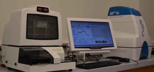
XF® 96 Extracellular Flux Analyzers are advanced cell metabolism analyzers
measuring two major energy producing pathways of cell-mitochondrial respiration
and glycolysis in a 96 well plate, in real-time. It can determine
in vitro oxygen consumption, glycolysis, CO2, mitochondrial dysfunction and fatty acid oxidation. Accompanying the
analyzer, is a Seahorse designed XF prep station to prepare culture plates
for assay; the prep station delivers fast, meticulous and thorough washing
of cells, ensuring consistent assay conditions.
Operating and Handling Procedures
Individual users design and run their own protocols, however design of
new protocols on the system must be done with core personnel. All reagents
and disposables must be provided by the user.
Data storage and retrieval:
All data are stored on an institutional server in a user specific folder;
the folder must be set up before the first use.
Data analysis:
Data analysis can be performed with the XF wave software manager installed
on the system computer as well as with software provided to investigators.
Data is also exportable to an Excel file.
Data Retention policy:
Data will be stored for 1 year from the collection date on the server.
Long-term storage is the responsibility of individual investigators.
Leica Laser MicroDissection (LMD7) Microscope System
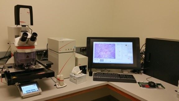
Laser Microdissection (LMD, also known as Laser Capture Microdissection or LCM) is typically used in genomics (DNA), transcriptomics (mRNA, miRNA), proteomics, metabolomics, and next generation sequencing (NGS). LMD enables users to isolate specific single cells or entire areas of tissue. Powered by a unique laser design and dynamic software, Leica LMD systems allow users to easily isolate Regions of Interest (ROI) from entire areas of tissue down to single cells or even subcellular structures such as chromosomes.
Operating and Handling Procedures
After completing training, researchers will be allowed to schedule time
on the system. Minor assistance will be provided as needed. Assistance
with analysis of data and other complex issues will be provided on a case-by-case basis.
Data storage and retrieval:
All data are stored on an institutional server in a user specific folder;
the folder must be set up before the first use.
Data analysis:
Data analysis is done with the Harmony software manager installed on the
system computer. Data is also exportable to an Excel file.
Data Retention policy:
Data will be stored for 1 year from the collection date on the server.
Long-term storage is the responsibility of individual investigators.
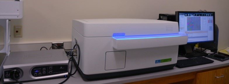
Perkin Elmer EnVision® Multilabel Reader
The EnVision® Multilabel Reader is a state-of-the-art dual-detector and fast high-throughput screening instrument.
Configuration:
Alpha technology: 570 nm
Absorbance: 405, 450, 540, 595 and 620 nm
Bioluminescence: 700 nm
Fluorescence assay: Excitation: 485 and 531 nm; Emission: 535 and 579 nm
Polarization assay for FITC is available.
Multilabel Plate Reader capabilities:
- Perkin Elmer Alpha technology analysis system
- Bioluminescence reader
- Fluorescence -based assay
- Multimodal assay
Operating and Handling Procedures
Users may schedule time on the EnVision Multilabel Reader through the online
scheduling system. All reagents and disposables must be provided by the user.
Data storage and retrieval:
All data are stored on an institutional server in a user specific folder;
the folder must be set up before the first use. Local data transfer can
be performed using storage devices such as USB or external hard drives.
Data analysis:
Data analysis is done with the Envision manager installed on the system
computer. Data is also exportable to an Excel file.
Data Retention policy:
Data will be stored for 1 year from the collection date on the server.
Long-term storage is the responsibility of individual investigators.
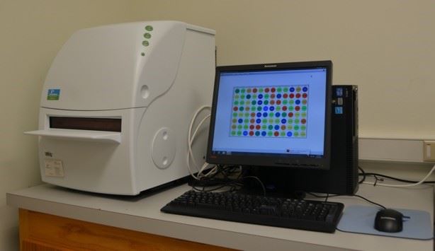
Agilent SureScan Microarray Scanner System
The SureScan Microarray Scanner is a state-of-art, laser-induced fluorescence
scanner designed to read microarrays printed on standard 1” ×
3” slides. The SureScan scanner measures the fluorescence intensity
of labeled sample nucleic acid (DNA and RNA) bound to microarrays. Data
extraction is handled by the Feature extraction program available on the
connected computer.
Scanner capabilities:
- Scan CGH, CGH+SNP, ChIP-on-chip, DNA methylation, microRNA arrays with dual color and up to 3 microns sensitivity
- Feature extraction software extracts data from scanned images.
Operating and Handling Procedures
Investigators design and run their own microarray slide hybridization protocols.
For hybridization of slides, the core houses a hybridization oven with
adjustable temperature. Scheduling to scan slides on the Agilent Surescan
Scanner is done through online scheduler. Slides can be scanned by core
personnel per the investigator protocol upon request.
Sample quality control:
DNA and RNA sample quality can be evaluated using either the BioRad Experion
or Agilent Bioanalyzer available in the core facility.
Data storage and retrieval:
All data are stored on an institutional server in a user specific folder;
the folder must be set up before the first use.
Data analysis:
Data extraction can be done with the Feature extraction software that is
provided on the system.
Data Retention policy:
Data will be stored for 1 year from the collection date on the server.
Long-term storage is the responsibility of individual investigators.
Molecular Devices Genepix 4100 A
The Genepix 4100A Microarray Scanner is a laser-induced fluorescence scanner
designed to read microarrays printed on standard 1" x 3" slides.
The Genepix scanner measures the fluorescence intensity of labeled sample
nucleic acids (DNA and RNA) and proteins bound to microarrays. Data extraction
is handled by the GenePix® Pro Microarray Analysis Software
available on the connected computer.
Scanner capabilities:
- Scan microarrays arrays with dual color, 532 and 635 nm excitation lasers and up to 5 microns sensitivity.
- GenePix™ Pro Microarray Analysis Software extracts data from scanned images.
Operating and Handling Procedures
Slide scanning and data extraction will be performed as required by core
personnel. If the researcher has more than 5 slides, they will be trained
on the system and then able to schedule time to scan and extract data
themselves. All reagents and disposables must be provided by the user .
Incucyte™ Live Cell Imaging System
This system automatically acquires live cell images, either fluorescence
(far red and green) or phase contrast images, inside a CO2 incubator in
multi-well plates and analyzes the data. An overview of all available
assays can be found Incucyte website.
Modules available in our core include: cell proliferation assay with or without dyes; cell migration assay by wound healing assay (wound maker available); chemotaxis assay; angiogenesis assay; spheroid growth assay; and Cell-by-Cell analysis software.
Operating and Handling Procedures
Training provided by core personnel upon request. There are 6 plate slots
available in a 3x2 rack set up; to avoid moving other investigators plates
only 3 researchers are allowed to use the system at a time. The system
can be booked up to 2 weeks in advance and is available on first come,
first served basis.
Data storage and retrieval:
All data are stored on an institutional server in a user specific folder;
the folder must be set up before the first use.
Data analysis:
Data analysis is done with the Harmony software manager installed on the
system computer. Data is also exportable to an Excel file.
Data Retention policy:
Data will be stored for 1 year from the collection date on the server.
Long-term storage is the responsibility of individual investigators.
Biorad CFX96™ Touch Real-Time PCR Detection System
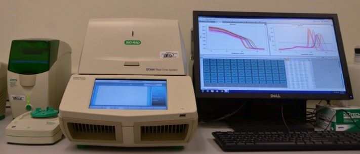
The CFX96™ Optical Reaction Module converts the C1000 Touch™ thermal cycler chassis into the powerful and precise CFX96 Touch Real-Time PCR Detection System.
Real-Time PCR capabilities:
- Calculation of the Ct values, melting temperature and quantification of gene expression
- CFX manager software
Operating and Handling Procedures
Individual users design and run their own protocols, however design of
new protocols on the system must be done with core personnel. All reagents
and disposables must be provided by the user. Use may be scheduled through
the online scheduler.
Data storage and retrieval:
All data are stored on an institutional server in a user specific folder;
the folder must be set up before the first use. Local data transfer can
be performed using storage devices such as USB or external hard drives.
Data analysis:
Data analysis is done with the CFX software manager installed on the system
computer. Data is also exportable to an Excel file. Free software (Windows
OS) is also available.
Data Retention policy:
Data will be stored for 1 year from the collection date on the server.
Long-term storage is the responsibility of individual investigators.
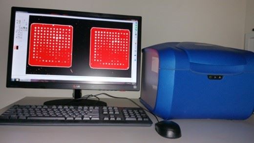
Oxford Optronix GelCount™ Colony Counter
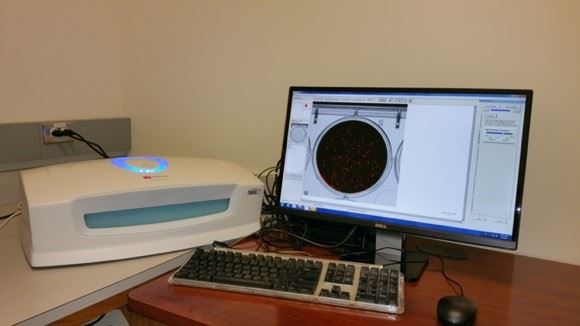
GelCount™ is an integrated, dedicated colony counter for processing tumour colony forming assays and is conceived to rapidly and accurately count as well as characterize mammalian cell colonies. Colonies can be adherent in nature (usually stained), or non-adherent, suspended either in liquid medium (spheroids) or in 3-dimensional semi-solid matrices such as soft agar and methylcellulose. Non-adherent cell colonies do not typically require staining.
Operating and Handling Procedures
After completing training, researchers will be allowed to schedule time
on the system. Minor assistance will be provided as needed. Assistance
with analysis of data and other complex issues will be provided on a case-by-case basis.
Data storage and retrieval:
All data are stored on an institutional server in a user specific folder;
the folder must be set up before the first use.
Data analysis:
Data analysis is done with the Harmony software manager installed on the
system computer. Data is also exportable to an Excel file.
Data Retention policy:
Data will be stored for 1 year from the collection date on the server.
Long-term storage is the responsibility of individual investigators.
Thermo Fisher iBright™
and MyECLGelimagers
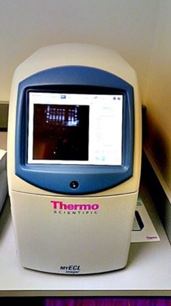
The iBright FL1500 Imaging System supports imaging of fluorescent-, chemiluminescent-, and colorimetric-stained western blots, fluorescent-stained nucleic acid gels, fluorescent- and colorimetric-stained protein gels, and colorimetric membrane stains.
My ECL™️ imager has the following acquisition modes: Chemiluminescence (Chemi) mode for image acquisition of self-emitting, chemiluminescent light; Ultraviolet (UV) mode for image acquisition of UV-transmitted light; Visible (Vis) mode for image acquisition of reflected and/or transmitted light
Operating and Handling Procedures
All reagents and disposables must be provided by the user.
Denovix Sphectrophotometer
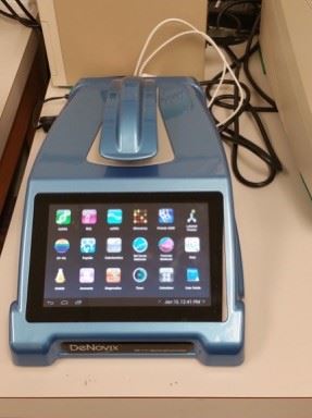
The DeNovix Spectrophotometer quantifies nucleic acids and protein using only 0.5 – 2.0 μL. Instant on-screen results include concentration, a full spectrum output, purity ratios and SmartQC™ contamination alerts.
Operating and Handling Procedures
Researchers may use the system during the core hours; no scheduling is required.
All reagents and disposables must be provided by the user.
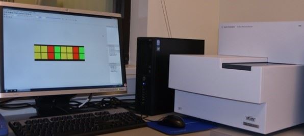
Biorad Experion™ Automated Electrophoresis Station and Agilent 2100
Bioanalyzer
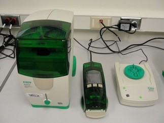
The Experion Automated Electrophoresis Station performs all steps for gel-based electrophoresis in one compact, durable unit. The Experion Automated Electrophoresis System can be used upstream and / or downstream of a number of nucleic acid- and protein-applications.
The Agilent 2100 Bioanalyzer is a microfluidics-based platform for sizing, quantification and quality control of DNA, RNA, proteins and cells.
Operating and Handling Procedures
Individual users design and run their own protocols, however design of
new protocols on the system must be done with core personnel. All reagents
and disposables must be provided by the user. Users may schedule through
the online scheduler.
Data storage and retrieval:
All data are stored on an institutional server in a user specific folder;
the folder must be set up before the first use. Local data transfer can
be performed using storage devices such as USB or external hard drives.
Data analysis:
Data analysis is done with the Experion manager or 2100 Expert installed
on their respective computers. Data is also exportable to an Excel file.
Data Retention policy:
Data will be stored for 1 year from the collection date on the server.
Long-term storage is the responsibility of individual investigators.
Nanoprint Microarray Printing
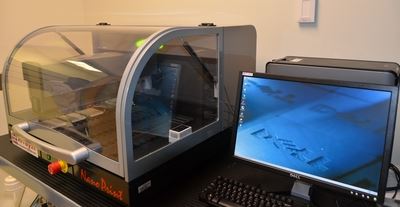
Nanoprint™ Microarrayers are platforms for all microarray manufacturing applications including DNA microarrays, protein microarrays, and other types of biomolecules. Based on advanced nanotechnology, Arrayit NanoPrint™ robots move with 500 nanometer resolution and deliver sub-nanoliter sample volumes using high throughput, patented contact printing devices.
Applications include:
- Reverse Phase protein lysate microarray
- Proteomic microarray
- Antibody array
- Peptide array
Operating and Handling Procedures
Microarray printing is provided as a service to the research community.
The protocol to use this facility is:
A project number is assigned to a proposed project; the project is discussed
with core personnel and a preliminary quote provided to the researcher.
Upon agreement on the quote, the required reagents will be procured. The
sample to be used for printing will be evaluated using appropriate quality
control protocols before printing.
Data storage and retrieval:
All data are stored on an institutional server in a user specific folder;
the folder must be set up before the first use.
Data Retention policy:
Data will be stored for 1 year from the collection date on the server.
Long-term storage is the responsibility of individual investigators.
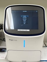
Qubit 4 Fluorometer
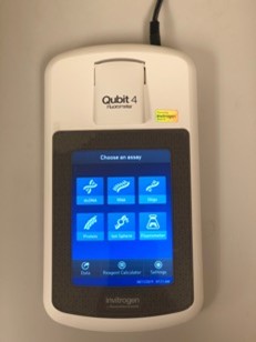
The Qubit 4 Fluorometer accurately measures DNA, RNA, and protein quantity, as well as RNA integrity and quality, using the sensitive Qubit assays. For all Qubit assays, the concentration or quality of the target molecule in the sample is reported by a fluorescent dye that emits a signal only when bound to the target, which minimizes the effects of contaminants on the result.
Key features of the Qubit 4 Fluorometer include:
- Fast and highly accurate quantitation of DNA, RNA, and protein in less than three seconds per sample
- Measures intact RNA in less than 5 seconds per sample
- High levels of accuracy using only 1–20 μL of sample, even with very dilute samples
- On-board Reagent Calculator quickly generates Qubit working solution preparation instructions.
Operating and Handling Procedures
Researchers use the system during core hours. No scheduling is required.
All reagents and disposables must be provided by the user.
Olympus Epifluorescence Inverted Microscope
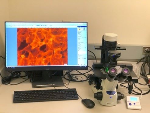
The Olympus inverted microscope is equipped with 4X, 10X and 20X objectives for viewing cells in cell culture dishes. There are 3 different fluorescence excitation and emission set ups available: DAPI, GFP and RFP. The microscope is equipped with a camera to capture images at low magnification.
Operating and Handling Procedures
Training provided by core personnel upon request.
Data storage and retrieval:
All data are stored on an institutional server in a user specific folder;
the folder must be set up before the first use.
Data analysis:
Data analysis is done with the Harmony software manager installed on the
system computer. Data is also exportable to an Excel file.
Data Retention policy:
Data will be stored for 1 year from the collection date on the server.
Long-term storage is the responsibility of individual investigators.
Thermo Fisher EVOS M7000 Imaging System
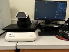
The EVOS M7000 Imaging System is a fully automatable, inverted, multi-channel fluorescence and transmitted light imaging system. It can be automated with enhanced autofocus algorithms and automated routines for microwell plate assays, time-lapse live cell imaging, area scanning in time lapse and Z-stacks in scan mode. The objectives available on the system are 4X, 20SXD, 20XLWD, 40X, and 40XLWD. The fluorescent channels available on the system are DAPI, Alexa488 and Alexa568. Images can be obtained from slides and 6- to 96-well format plates.
Operating and Handling Procedures
Training provided by core personnel upon request.
Data storage and retrieval:
All data are stored on an institutional server in a user specific folder;
the folder must be set up before the first use.
Data analysis:
Data analysis is done with the Harmony software manager installed on the
system computer. Data is also exportable to an Excel file.
Data Retention policy:
Data will be stored for 1 year from the collection date on the server.
Long-term storage is the responsibility of individual investigators.
Perkin Elmer Janus™ Automated Workstation
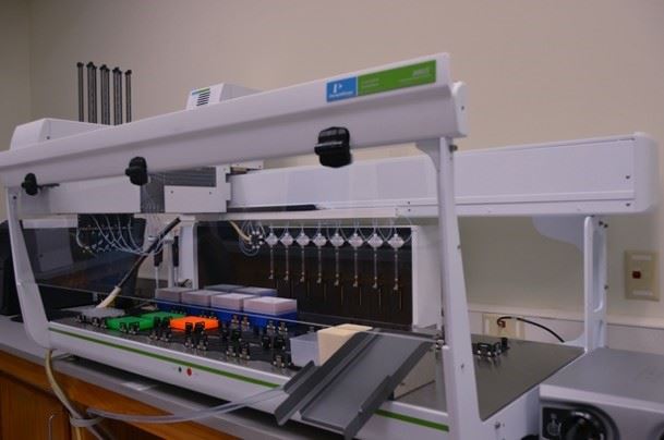
The Janus™ Automated Workstation handles liquid in the hands-off “on the fly” adaptability fashion. It provides precision pipetting from nanoliters to microliters in seconds for up to 1536-well plates. The workstation is used to set up high throughput assays, in combination with other equipment.
Applications include:
- High throughput DNA/RNA extraction using magnetic beads
- Set up deep sequencing plate with consistent pipetting
- Set up real-time PCR plates automated for precision pipetting
- Serial dilutions to achieve absolute concentrations during pharmacological screening of compounds
The core is available to work with investigators to develop any individualized high throughput assay program.
Operating and Handling Procedures
Liquid handling by the Janus automated workstation must be programmed and
tested for any high throughput assay set up. A project number is assigned
and the assay discussed in detail with the investigator. The program will
be built and tested before executing the assay itself. Trained users for
specific programs will be allowed to schedule the equipment for the required
program execution time. All reagents and disposables must be provided
by the user.
Rarecell ISET Circulating Cells
ISET®, Isolation by SizE of Tumor Cells, is based on the antibody-independent
whole blood filtration approach for circulating tumor cell (CTC) and micro-emboli
isolation. The technique relies on the larger size of all types of CTC
as compared to leukocytes. The ISET system successfully captures CTC that
are presenting, by definition, heterogeneous phenotypic profiles.
Simple workflow:
10 mL blood draw collected in EDTA or STRECK tubes,
Sample mixed with application-specific buffer for integrity of CTC structure
Sample processed on the ISET© Device pressure-controlled filtration system
Fixed cells: Multiplexed downstream processing from the 10 spots on the membrane
Operating and Handling Procedures
This system can be used in the Principal Investigator’s laboratory.
An approved IRB protocol is required before use of the system is permitted.
Data storage and retrieval:
All data are stored on an institutional server in a user specific folder;
the folder must be set up before the first use.
Data analysis:
Data analysis is done with the Harmony software manager installed on the
system computer. Data is also exportable to an Excel file.
Data Retention policy:
Data will be stored for 1 year from the collection date on the server.
Long-term storage is the responsibility of individual investigators.